
Subtopic 2.3: Structure of Chromatin
Chromosomal DNA is packaged inside microscopic nuclei by its association of histones H2A, H2B, H3, and H4. These are positively-charged proteins that strongly adhere to negatively-charged DNA and form complexes called nucleosomes. (11-nm “beads on a string” structure). Each nucleosome is composed of DNA wound 1.65 times around eight histone proteins. Nucleosomes fold up to form a 30-nanometer chromatin fiber, which forms loops averaging 300 nanometers in length. The 300 nm fibers are compressed and folded to produce a 250 nm-wide fiber, which is tightly coiled into the chromatid of a chromosome. The level of structure varies depending on the cell cycle stage and, as a result, the requirement for DNA transcription or replication.
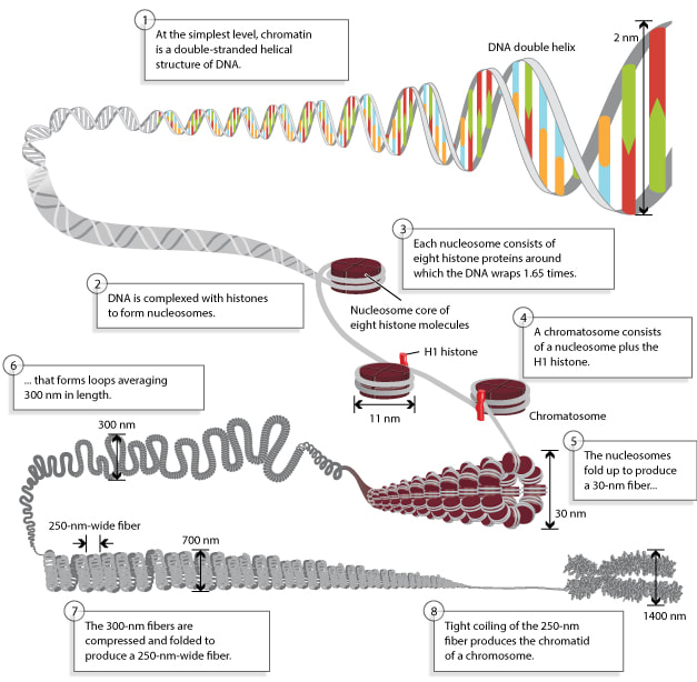
Figure 3: Chromosomes are composed of DNA tightly-wound around histones - © 2010 Nature Education
Chromatin: The most important structure inside the nucleus is chromatin, consisting, in humans, of the 46 chromosomes. Chromatin is the combination or complex of DNA and proteins that make up the contents of the nucleus of a cell. The most abundant protein in the nucleus are histones. Histones are rich in basic amino acids (positively charged), which interact with negative charges of the DNA.

|

|
|
|